23+ Plant Cell Under Microscope
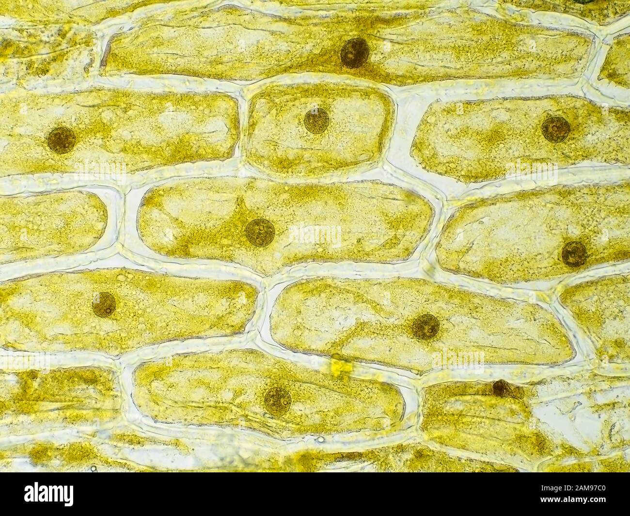
Figure 5 Making wet preparations yourself Figure 7 Work on the ZEISS Primo Star microscope Figure 8 Hand drawing Figure 6 Epidermis cell of the onion skin under the microscope phase contrast The epidermal cells of the onion display a structured cell. Learn about the cell wall nucleus chloroplasts and vacuoles.

Plant Cell Images Plant Cell Plant Tissue
The cell wall is distinctly visible around each cell.
. Web We present a new large-scale three-fold annotated microscopy image dataset aiming to advance the plant cell biology research by exploring different cell microstructures including cell size. Web It was not until good light microscopes became available in the early part of the nineteenth century that all plant and animal tissues were discovered to be aggregates of individual cells. A diagram of a plant cell.
Cell is a tiny structure and functional unit of a living organism containing various parts known as organelles. Most photographs of cells are taken using a microscope and these pictures can also be called micrographs. Web There is an enormous suite of microscopy tools available to the cell biologist from the simplest bright-field light microscopy to the highly technical transmission electron microscopy TEM.
Camera - Nikon D3300. Plant cell under the microscope. Humans have been making use of plants for thousands of years.
Web The past decades have seen a revolution in microscopy approaches to study cellular biology mostly in the fields of live-cell imaging using fluorescence microscopy three-dimensional electron microscopy 3D-EM and correlative light and electron microscopy CLEM. Web Learn the structure of animal cell and plant cell under light microscope. Below we review how live imaging has taken on central importance in the emergence of this field of cell and developmental biology.
Web Microscopy and stained specimens engage students visually as they learn about plant anatomy a topic covered in many biology and introductory science courses. Web for the preparation. Cells are the smallest part of a living organism and are around 001 mm - 003 mm long.
See how a generalized structure of an animal cell and plant cell look with labeled diagrams. Fluorescence-based light microscopy allows us to visualize. Web A microscope is an instrument that magnifies objects otherwise too small to be seen producing an image in which the object appears larger.
Web When the researchers looked through the microscope the miniature world of plant cells and organelles was brought into sharp focus. It goes further to explain and demonstrate the plant cell and animal cell as seen under both the light and electron microscope. Comments are turned off.
Web Calculating magnification Key points All living organisms are made up of cells. Look at the slide with your microscopes 10x objective to see the general structure and higher power to see cell. Web New imaging methodologies with high contrast and molecular specificity allow researchers to analyze dynamic processes in plant cells at multiple scales from single protein and RNA molecules to organelles and cells to whole organs and tissues.
The nucleus and chloroplasts of eukaryotic cells can also be seenhowever smaller organelles and viruses are beyond the limit of resolution of the light microscope see Figure 1. To look at a cell close. Web Biologists typically use microscopes to view all types of cells including plant cells animal cells protozoa algae fungi and bacteria.
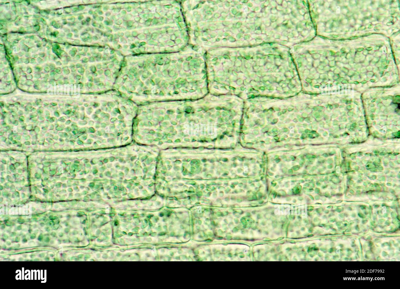
Web This video tries to distinguish between a cell and an organelle. Plants cells differ from animal cells in that they have a cell wall which is glued to adjacent cells by the middle lamellae a large central vacuole and chloroplasts. Under the microscope plant cells are seen as large rectangular interlocking blocks.
I used the aquatic plant anacharis Egeria densa and a Marimo ball Aegagropila linnaei. You can do this by adding a drop or two of water over the leaf section and then covering it with the coverslip. In this activity students section plant material and prepare specimens to view under a brightfield microscope.
This discovery proposed as the cell doctrine by Schleiden and Schwann in 1838 marks the formal birth of cell biology. The cell wall is somewhat thick and is seen rightly when stained. 18 June 2021 Biosensor imaging of a seedling measuring how the concentrations of the plant hormone gibberellin change as the plant grows.
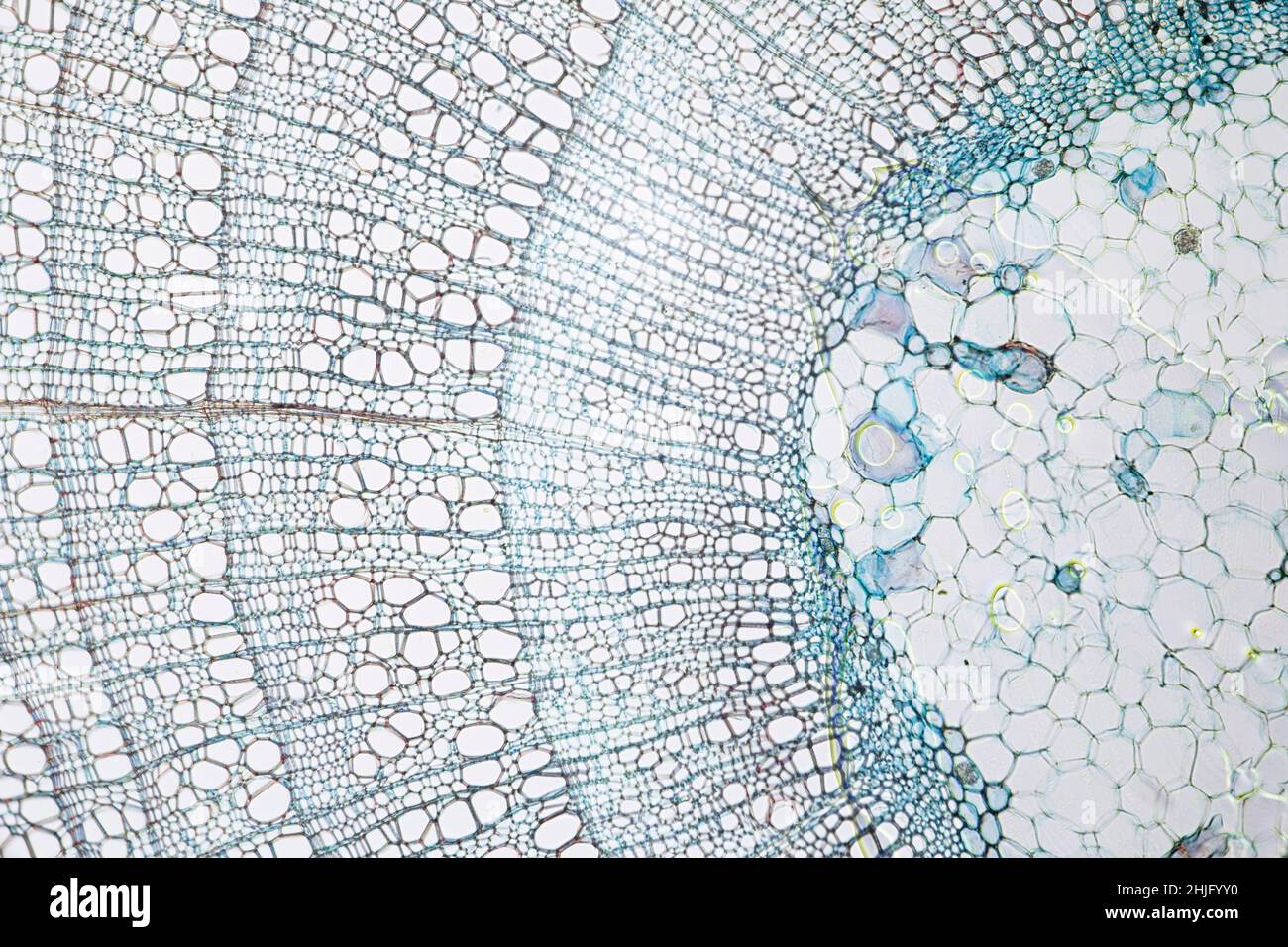
By looking at the microscopic structures of different parts of the plant parts we can learn how the plant function at the cellular level. Under the microscope the structure of a plant cell becomes visible. Web Plant cells under the microscope Science Skool 42K subscribers Subscribe 26K views 4 years ago A short video showing the cells of plants and how they may look under the.
Explore what each of these parts does in the cell. Each part has its unique job to keep the whole plant healthy. Web In the last decades biophysical approaches have been developed to probe the mechanical properties of plant cells and their response to forces.
What you see when looking at an elodea leaf under a microscope. Web Plant Cells Under a Microscope. Web Make a wet mount on a plain slide with the inner part of the leaf section facing up so the inner cells are visible.
Discover the parts of a plant cell and what differentiates it from the cells of other organisms. Web Sped up microscopic footage of oxygen bubbles in water produced from photosynthesis. The rigid lines of cell walls stood out in bas-relief from the.
Using a camera or cell phone images of microscope slide. Web The five main parts are the roots the leaves the stem the flower and the seed.
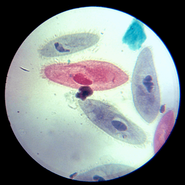
Amazing 27 Things Under The Microscope With Diagrams

4 971 Plant Cell Under Microscope Images Stock Photos Vectors Shutterstock

6 700 Plant Cell Microscope Stock Photos Pictures Royalty Free Images Istock Plant Cell Wall

Komorki Epidermy Plant Cell Things Under A Microscope Plant Cell Picture
How To See A Plant Cell Under A Compound Microscope Quora
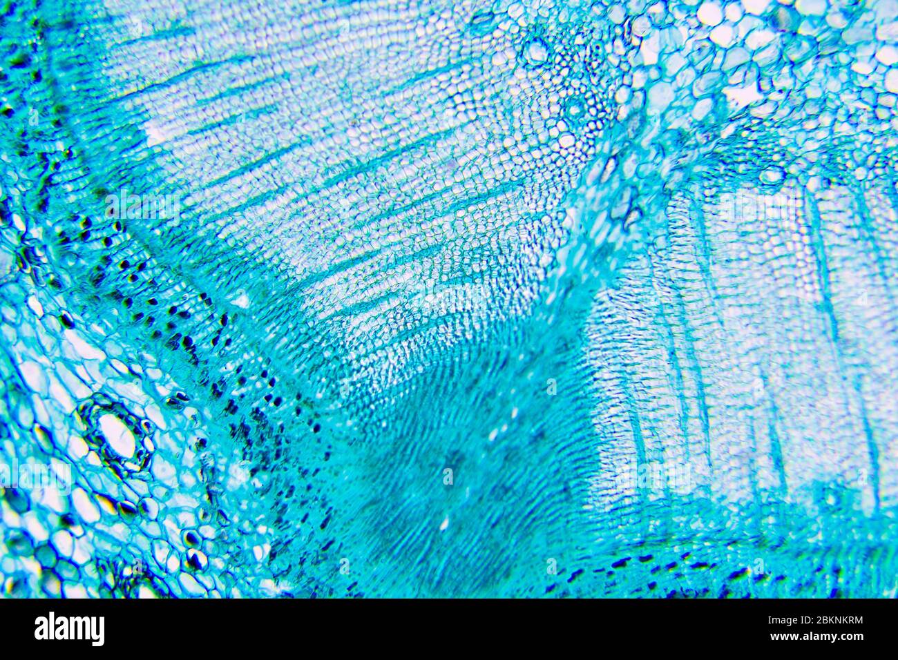
Plant Cells Under Microscope Stock Photo Alamy
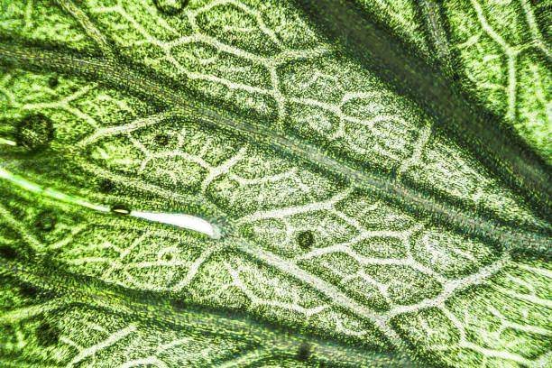
6 700 Plant Cell Microscope Stock Photos Pictures Royalty Free Images Istock Plant Cell Wall
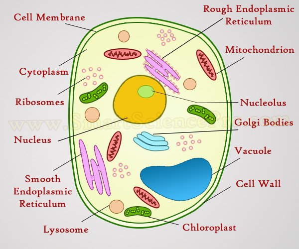
Structure Of Animal Cell And Plant Cell Under Microscope Diagrams
Plant Cell Microscope Hi Res Stock Photography And Images Alamy

4 971 Plant Cell Under Microscope Images Stock Photos Vectors Shutterstock
Plant Cells Microscopic Hi Res Stock Photography And Images Alamy
Plant Cells Microscope Hi Res Stock Photography And Images Alamy
Amazing 27 Things Under The Microscope With Diagrams

Plant Cell Under The Microscope 1 프랙털 나뭇잎

4 971 Plant Cell Under Microscope Images Stock Photos Vectors Shutterstock
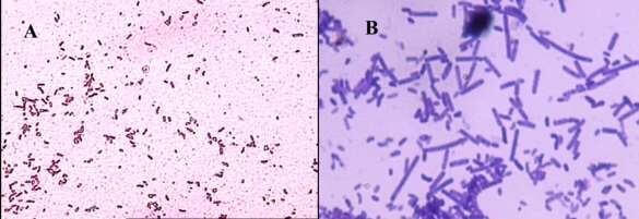
Amazing 27 Things Under The Microscope With Diagrams

277 Plant Cells Under Microscope Stock Photos High Res Pictures And Images Getty Images